
Tissue engineering is a multidisciplinary sector of biotechnologies which paves the way for new treatment options and better quality of life for patients. Tissue engineering techniques entail the use of biomaterials and scaffolds that must meet specific requirements and certain standards. Through the use of micro computerised tomography (microCT) it is possible to obtain a wealth of information on the internal structure of scaffolds and biomaterials, providing high resolution images without damaging the sample and with no need for cuts, lining or chemical treatments in preparation for the test. MicroCT is therefore a tool of fundamental importance in Medicine, Biology and Biomedical Engineering, and makes it possible to carry out thorough and detailed 3D morphological analyses, structural property assessments, multi-phase characterisations, identification and quantification of defects in biomaterials and scaffolds.
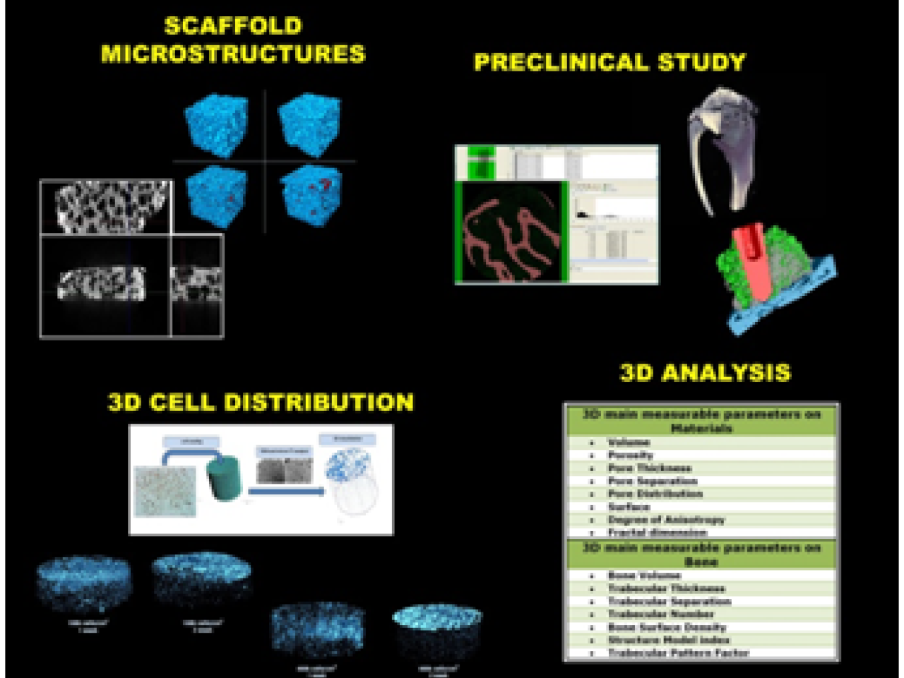 Main applications of the service offered
2D histomorphometry techniques are not able to offer a complete picture of the bone's three-dimensional micro-structure. Starting from micro-tomographic sections, however, it is possible to create virtual 3D models of the object being analysed, enabling realistic spatial visualisations of the micro-structures, providing an immediate and interactive assessment of the devices and their operation. The models may be used to create videos of the object being analysed in motion or as a basis for prototypes in tissue engineering
The product obtained will therefore be able to meet the highest quality standards and be launched on the market after strict and highly competitive assessment, which includes a wide number of tests, both qualitative and quantitative. MicroCT testing may also be applied to any type of material used, not only biomedical, providing both micro-structural and mechanical three-dimensional information.
 Tissue engineering 3D models
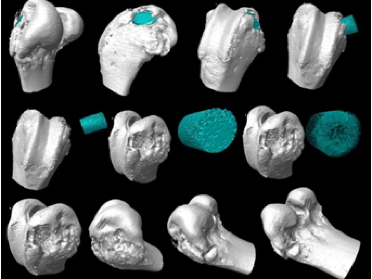
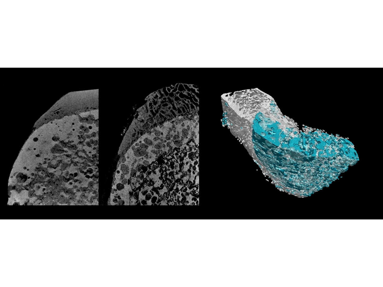
Assessment of cranial implants based on porous hydroxyapatite
The product allowed a hydroxyapatite-based cranial bone substitute to be assessed by means of three-dimensional analysis, through the evaluation of microstructural parameters such as volume of the pores, connection, separation, thickness and, finally, spatial distribution, elements that cannot be analysed by two-dimensional static and/or dynamic analysis. Following these results 3D analysis has also been used to assess hydroxyapatite-based cranial bone substitutes in vivo and from peri-implant bone biopsies. These assessments have made it possible to obtain information concerning the implanted material and the surrounding bone following extensive losses of substance, resulting, for example, from severe congenital malformations, traumatic events and neoplastic lesions. The results obtained from three-dimensional assessments through microCT have made it possible to detect micro-structural changes in porosity of cranial bone substitutes after being in contact with biological fluids and tissue. The data obtained have been fundamentally important in understanding the dynamic interaction between scaffold and bone, affording a comprehensive vision of the micro and macro-structure both of the implanted material and the surrounding bone.
Preclinical and Surgical Studies Laboratory, Codivilla Putti Research Institute, Rizzoli Orthopaedics Institute Fin-Ceramica Faenza S.p.A
Translation and clinical application of the tested innovative bio-materials and scaffolds
 Translation and clinical application of the tested innovative bio-materials and scaffolds