
Modern 'light sheet' microscopy techniques combined with three-dimensional image processing and analysis using high-powered computing media enable the visualisation of microscopic structures from an entire organ or even an entire lab mouse. This is possible by sample preparation procedures that make the sample transparent to laser light ('clarification'). The published methods involve the use of highly toxic organic solvents, in protected environments during both the clarification and the visualisation of the sample, and the disposal of residual reagents as special waste. IRET Foundation, in collaboration with Miltenyi Biotec, has developed a complete workflow using non-toxic Miltenyi products, optimized to date for visualisation and quantification by 'voxel-based' image analysis of the entire cerebral vascular system and axon structures both in the brain and in the spinal cord.
 IRET Foundation, the managing body of the Bologna Technopole, Ozzano dell'Emilia 'Rita Levi-Montalcini' site
The developed workflow includes the sample preparation procedure optimized to date for brain and spinal cord through a clarification procedure using the 'MACS® Clearing Kit' from Miltenyi Biotec, which can be performed in a conventional laboratory environment with a few steps under a normal chemical fume hood. The observation of the sample immersed in Miltenyi Biotec's 'MACS® Imaging Solution' using the UltraMicroscope Blaze™ is also conducted in a conventional environment. UltraMicroscope Blaze™ is equipped with 1×, 4× and 12× objectives, with 'magnification lenses' up to a maximum of 36×, allowing acquisition at different levels of cellular and subcellular resolution (0.5 µm). The images acquired (datasets) by the Blaze, are in the size range of giga/tera bytes and are managed by a dedicated workstation. The final stage of the procedure involves the use of image analysis software based on the constituent voxels of the image itself, enabling image quantification procedures (also assisted by machine learning/AI algorithms) from fluorescence intensity.
This microscopy, when optimized, allows the visualization and analysis of macroscopic samples with cellular resolution. It not only provides a three-dimensional view of the histological organization of organs and tissues, but also allows quantitative evaluation. It can be used to visualize endogenous fluorescent signals (e.g. fluorescence genetically encoded proteins), but also with single antibodies or in combination.
The Light Sheet technology ‘UltraMicroscope Blaze’ can be used for both basic and translational research. It therefore allows the robust and repeatable analysis of healthy and pathological samples, and its use is also promising in the effectiveness investigation of drug and non-drug therapies. Compared to 2D microscopic techniques, it allows a more rapid analysis of all areas of interest within the organ being analyzed.
Compared to currently used clarification techniques (iDISCO, 3DISCO, etc.), the used workflow employs non-toxic solvents and is therefore fully in line with the DNSH (Do No Significant Harm) principle introduced by EU Regulation 852/2020.
The applications that have been developed to date include:
1. Study of the cerebral vascular bed, which can be visualized both by administering tracers such as dextran-FITC and by using antibodies for endothelial targets (CD31). The simultaneous visualisation of dextran-FITC and CD31 allows imaging and quantification of the entire circulatory system, arteries and veins down to the smallest capillaries (3-4 µm diameter);
2. Study of the cerebral and spinal pathways, using anti-neurofilament (NF) antibody for the study in injury models (e.g. spinal cord contusion injury).
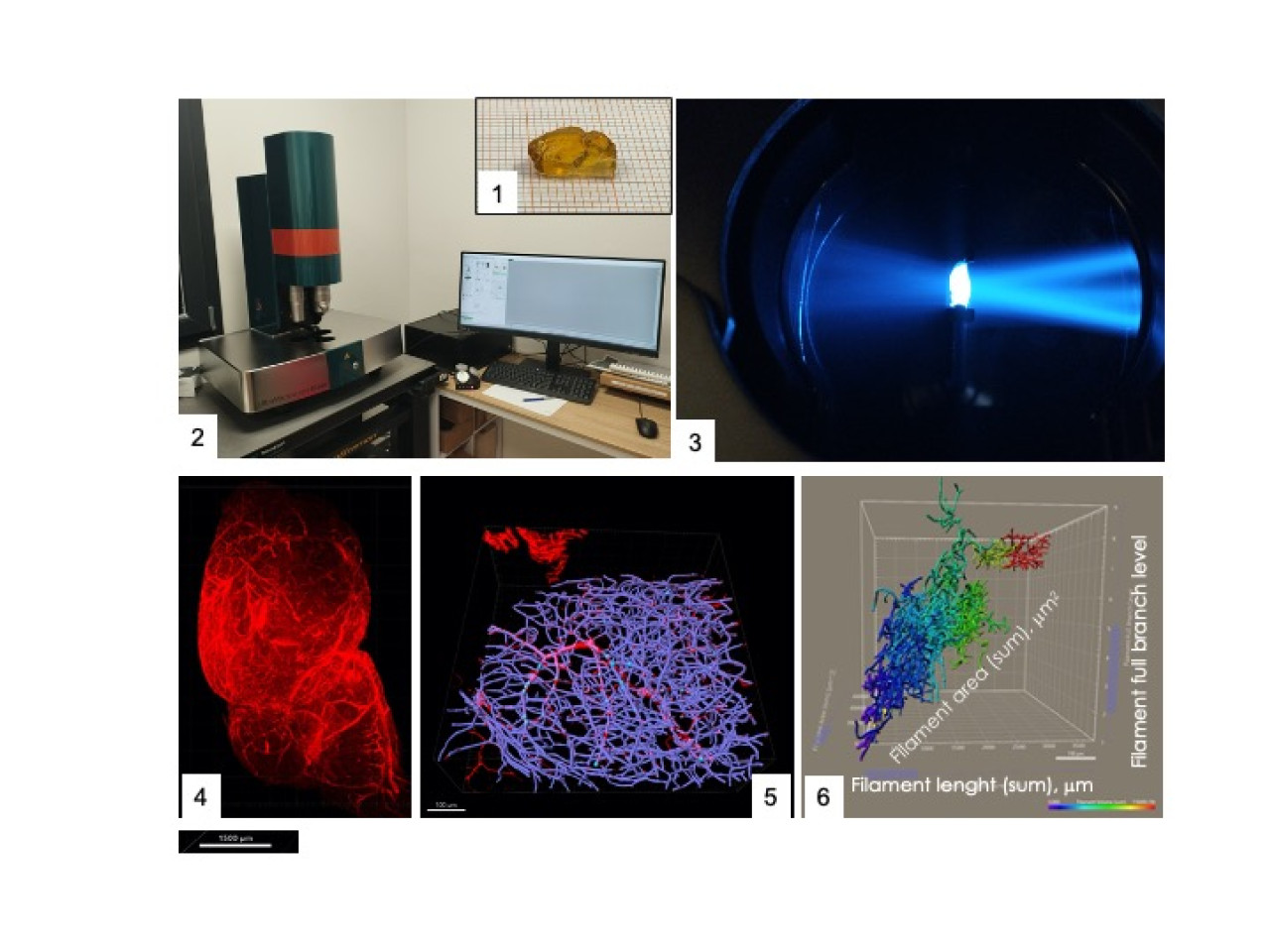 Working flowchart for three-dimensional visualisation of large tissue samples. 1. clarification; 2-3. acquisition; 4. visualisation; 5. image post processing; 6. quantification
The presented workflow can be applied to:
- Basic studies for a better understanding of the histological organization of organs and tissues
- Translational research for the study of structural alterations in animal models
- Pharmacological research for studying the effectiveness of innovative treatments
- Development of new algorithms for three-dimensional image analysis
Structural and functional alterations in the microcirculation are recognized as a primary pathogenetic factor in many degenerative diseases. The illustrated procedure was applied to the study of the microcirculation in mice models of Alzheimer's disease with the aim of defining its temporal evolution, from the asymptomatic phase to the overt phase of the disease defined by the presence of the characteristic histopathological alterations (amyloid plaques) and the cognitive defect (learning and memory).
The different brain areas were analyzed, defining a three-dimensional map including the different cortical areas, hippocampal regions and cerebellum. This map is the basis for superimposing histological but also spatial transcriptomic-derived images, thus enabling a correlation between microcirculation in tissue volumes and expression of genes of interest. The results will be the subject of scientific publication in an international journal.
Miltenyi Biotec S.r.l., Italy
IRET Foundation, the managing body of the Bologna-Ozzano Technopole, together with Miltenyi Biotec, organized three ‘Blazing Days’ events in 2023, which are focused on presenting the complete workflow (products and tool). The last event (Nov. 2023) was dedicated to the winners of the Creative BraYns award, organised by the BraYn Association.
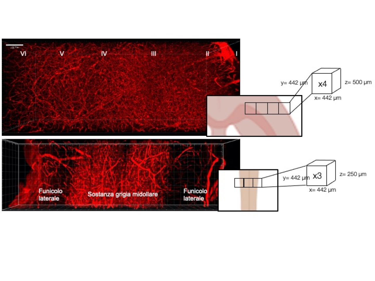 Visualisation of CD31 in 'whole hemibrain and spinal cord' of mice. The diagrams on the right show the sampling strategy and dimensions of the final 3D image.